Over the past decade, robotic neurorehabilitation has become one of the most discussed innovations in neurological recovery. Robotic gait trainers, upper-limb rehabilitation systems, exoskeletons, and AI-assisted rehabilitation devices are increasingly being adopted by hospitals and rehabilitation centres worldwide. However, an important question remains: Are robots the future of neurorehabilitation—or are they simply another tool in the rehabilitation toolbox? As clinicians and researchers, we must move beyond marketing claims and focus on scientific evidence, patient selection, and clinical reasoning. What is Robotic Neurorehabilitation? Robotic neurorehabilitation involves the use of electromechanical devices that assist, guide, resist, or augment movement during therapy. These technologies include: • Robotic gait trainers • Wearable exoskeletons • Upper limb robotic rehabilitation devices • End-effector robotic systems • Sensor-based rehabilitation platforms • AI-assiste...

Electroencephalography (EEG)
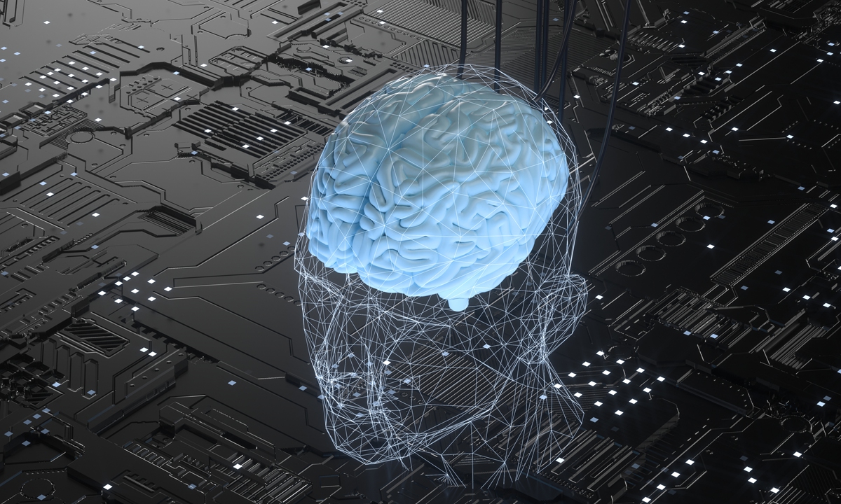
Based Brain-Computer Interfaces (BCIs) are systems that enable communication
and control of external devices directly through brain activity, measured via
electrodes placed on the scalp.
1. Overview of EEG Technology
Electroencephalography
(EEG) is a widely used, non-invasive technique for recording
electrical activity in the brain. EEG captures the electrical impulses produced
when neurons communicate, providing insights into brain state and function.
1.1 Principles
of EEG
- Electrical Signaling:
Neurons generate electrical signals when they fire, and groups of neurons
produce synchronized electrical activity that can be detected on the scalp
through electrodes.
- Signal Detection:
EEG electrodes measure voltage fluctuations resulting from ionic current
flows within the neurons, reflecting the brain’s electrical activity in
terms of rhythms (e.g., alpha, beta, delta, and theta waves).
2. Mechanisms of EEG-Based BCI
2.1 Data
Acquisition
- Electrode Placement:
Electrodes are typically placed on the scalp following standardized
configurations, such as the 10-20 system, to ensure consistent and
reproducible recording locations.
- Signal Amplification:
The tiny voltage signals picked up by the electrodes are amplified for
better quality before processing.
2.2 Signal
Processing
- Preprocessing:
Raw EEG data undergoes filtering to reduce noise and artifacts (e.g., from
eye movements, muscle contractions, or external electrical interference).
- Feature Extraction:
Significant features are extracted from the processed signals to represent
the user's intentions or mental states. Common features include
event-related potentials (ERPs), spectral power features (e.g., alpha and
beta band power), or time-domain features.
2.3
Classification and Control
- Machine Learning Algorithms:
Extracted features are used to train machine learning models that classify
brain states or user intentions. Common classification techniques include
support vector machines (SVM), neural networks, and linear discriminant
analysis (LDA).
- Control Mechanism:
The classified outputs are translated into commands that control external
devices, such as a cursor on a screen, robotic limbs, or other assistive
technology.
3. Applications of EEG-Based BCIs
3.1
Communication for Individuals with Disabilities
- Assistive Communication Devices:
EEG BCIs enable users with severe motor impairments (e.g., Amyotrophic
Lateral Sclerosis, locked-in syndrome) to communicate through direct
thought processes, allowing them to select letters or words.
3.2 Control of
External Devices
- Neuroprosthetics and Robotics:
EEG BCIs are used to control robotic arms or wheelchairs, allowing users
to perform tasks through thought alone, improving independence and quality
of life.
3.3 Cognitive
and Mental State Monitoring
- Cognitive Load and Attention Tracking:
EEG can be applied in workplace or educational environments to monitor
cognitive load, fatigue, and attention levels, helping optimize task
performance or training programs.
4. Advantages of EEG-Based BCIs
4.1 Non-Invasive
and Safe
- EEG technology is safe and does not require invasive
procedures, making it suitable for long-term use and repeated applications
without health risks.
4.2 Real-Time
Data Acquisition
- EEG provides near real-time monitoring of brain activity,
allowing for instantaneous feedback and control, which is critical for
applications requiring quick responses.
4.3
Cost-Effective
- EEG systems are generally less expensive than other
neuroimaging technologies, such as fMRI or MEG, making them more
accessible for research and clinical environments.
5. Challenges and Limitations
5.1 Spatial
Resolution
- The spatial resolution of EEG is relatively low compared
to other imaging techniques like fMRI, as it primarily reflects surface
cortical activity rather than deeper brain structures.
5.2 Noise and
Artifacts
- EEG signals are susceptible to various artifacts,
including those from eye movements (e.g., blink artifacts), muscle
activity, and electrical interference, which can complicate data
interpretation.
5.3 Variability
Across Subjects
- Individual differences in brain structure and function
can lead to variability in EEG signals, making it challenging to develop
universal BCI systems applicable to diverse populations.
6. Future Directions for
EEG-Based BCIs
6.1 Hybrid
Systems
- Research into hybrid systems that combine EEG with other
technologies (e.g., fNIRS, fMRI) may enhance spatial and temporal
resolution, providing comprehensive insights into brain activity.
6.2 Advanced Machine
Learning Techniques
- Continuous advancements in machine learning and
artificial intelligence can improve the accuracy and reliability of EEG
signal classification, making BCIs more efficient and user-friendly.
6.3 Clinical
Advancements
- Further research into EEG-based BCIs has the potential to
revolutionize rehabilitation strategies for neurological disorders such as
stroke, traumatic brain injury, or neurodegenerative diseases, offering
new avenues for patient recovery.
Conclusion
EEG-based Brain-Computer
Interfaces provide an innovative means for facilitating communication and
control through direct interaction with brain activity. With advantages such as
being non-invasive, cost-effective, and capable of real-time data acquisition,
EEG technology holds tremendous potential for enhancing the quality of life for
individuals with disabilities and expanding our understanding of cognitive
processes. Despite challenges regarding spatial resolution and susceptibility
to artifacts, ongoing advancements and research into hybrid solutions and
machine learning techniques will likely shape the future of EEG-based BCIs,
paving the way for practical applications across clinical, educational, and
entertainment domains.
Comments
Post a Comment