
Development of Prefrontal Cortex: Regions of PFC


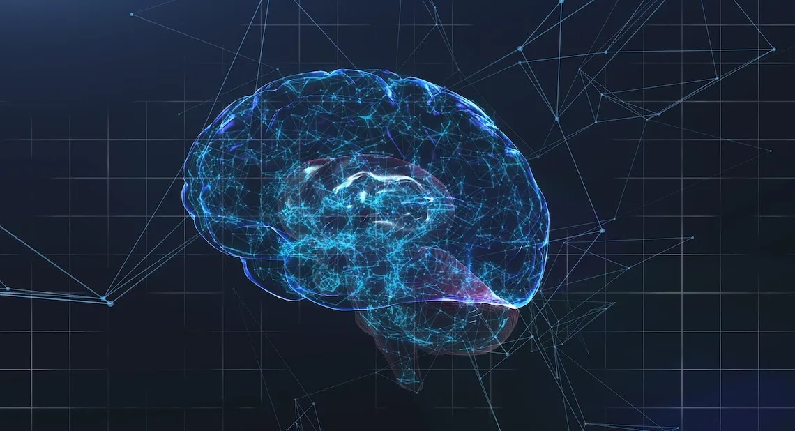
The prefrontal cortex (PFC) is a critical brain region associated with higher-order cognitive functions, including executive function, decision-making, social behavior, and emotional regulation. The PFC undergoes significant development across the lifespan, with distinct regions contributing to various aspects of cognitive control and behavior. Here are the key regions of the prefrontal cortex and their functions:

1.     Orbitofrontal Cortex (BA 11):

o    Location: Located in the ventromedial part of the PFC.

o  Function: The orbitofrontal cortex is involved in decision-making, reward processing, emotional regulation, and social behavior. It plays a role in evaluating the emotional and motivational significance of stimuli and guiding adaptive behavior based on reward outcomes.

2.     Ventrolateral PFC (BA 44, 45, 47):

o    Location: Situated in the lower lateral aspects of the PFC.

o Function: The ventrolateral PFC is associated with cognitive control, working memory, language processing, and response inhibition. It plays a role in maintaining task-relevant information, manipulating information, and regulating attention during complex cognitive tasks.

3.     Dorsolateral PFC (BA 9, 46):

o    Location: Located in the upper lateral aspects of the PFC.

o   Function: The dorsolateral PFC is involved in executive functions such as planning, problem-solving, cognitive flexibility, and goal-directed behavior. It plays a crucial role in working memory, mental manipulation of information, and strategic decision-making.

4.     Rostrolateral PFC (BA 10):

o    Location: Situated in the rostral part of the lateral PFC.

o Function: The rostrolateral PFC is associated with cognitive control, attentional processes, and multitasking. It plays a role in monitoring and coordinating complex cognitive operations, integrating information from multiple sources, and maintaining task sets for goal-directed behavior.

These regions of the prefrontal cortex work in concert to support various aspects of executive function and cognitive control. The hierarchical organization of the PFC allows for the integration of information, the regulation of behavior, and the coordination of complex cognitive processes. Understanding the functions of different regions within the prefrontal cortex provides insights into the neural basis of higher cognitive functions and the role of the PFC in adaptive behavior and decision-making processes.


Comments

Popular posts from this blog

Research Process

The research process is a systematic and organized series of steps that researchers follow to investigate a research problem, gather relevant data, analyze information, draw conclusions, and communicate findings. The research process typically involves the following key stages: Identifying the Research Problem : The first step in the research process is to identify a clear and specific research problem or question that the study aims to address. Researchers define the scope, objectives, and significance of the research problem to guide the subsequent stages of the research process. Reviewing Existing Literature : Researchers conduct a comprehensive review of existing literature, studies, and theories related to the research topic to build a theoretical framework and understand the current state of knowledge in the field. Literature review helps researchers identify gaps, trends, controversies, and research oppo...

Mglearn

mglearn is a utility Python library created specifically as a companion. It is designed to simplify the coding experience by providing helper functions for plotting, data loading, and illustrating machine learning concepts. Purpose and Role of mglearn: ·          Illustrative Utility Library: mglearn includes functions that help visualize machine learning algorithms, datasets, and decision boundaries, which are especially useful for educational purposes and building intuition about how algorithms work. ·          Clean Code Examples: By using mglearn, the authors avoid cluttering the book’s example code with repetitive plotting or data preparation details, enabling readers to focus on core concepts without getting bogged down in boilerplate code. ·          Pre-packaged Example Datasets: It provides easy access to interesting datasets used throughout the book f...

Distinguishing Features of Vertex Sharp Transients

Vertex Sharp Transients (VSTs) have several distinguishing features that help differentiate them from other EEG patterns.  1.       Waveform Morphology : §   Triphasic Structure : VSTs typically exhibit a triphasic waveform, consisting of two small positive waves surrounding a larger negative sharp wave. This triphasic pattern is a hallmark of VSTs and is crucial for their identification. §   Diphasic and Monophasic Variants : While triphasic is the most common form, VSTs can also appear as diphasic (two phases) or even monophasic (one phase) waveforms, though these are less typical. 2.      Phase Reversal : §   VSTs demonstrate a phase reversal at the vertex (Cz electrode) and may show phase reversals at adjacent electrodes (C3 and C4). This characteristic helps confirm their midline origin and distinguishes them from other EEG patterns. 3.      Location : §   VSTs are primarily recorded from midl...

Distinguishing Features of K Complexes

  K complexes are specific waveforms observed in electroencephalograms (EEGs) during sleep, particularly in stages 2 and 3 of non-REM sleep. Here are the distinguishing features of K complexes: 1.       Morphology : o     K complexes are characterized by a sharp negative deflection followed by a slower positive wave. This biphasic pattern is a key feature that differentiates K complexes from other EEG waveforms, such as vertex sharp transients (VSTs). 2.      Duration : o     K complexes typically have a longer duration compared to other transient waveforms. They can last for several hundred milliseconds, which helps in distinguishing them from shorter waveforms like VSTs. 3.      Amplitude : o     The amplitude of K complexes is often similar to that of the higher amplitude slow waves present in the background EEG. However, K complexes can stand out due to their ...

Maximum Stimulator Output (MSO)

Maximum Stimulator Output (MSO) refers to the highest intensity level that a transcranial magnetic stimulation (TMS) device can deliver. MSO is an important parameter in TMS procedures as it determines the maximum strength of the magnetic field generated by the TMS coil. Here is an overview of MSO in the context of TMS: 1.   Definition : o   MSO is typically expressed as a percentage of the maximum output capacity of the TMS device. For example, if a TMS device has an MSO of 100%, it means that it is operating at its maximum output level. 2.    Significance : o    Safety : Setting the stimulation intensity below the MSO ensures that the TMS procedure remains within safe limits to prevent adverse effects or discomfort to the individual undergoing the stimulation. o Standardization : Establishing the MSO allows researchers and clinicians to control and report the intensity of TMS stimulation consistently across studies and clinical applications. o   Indi...