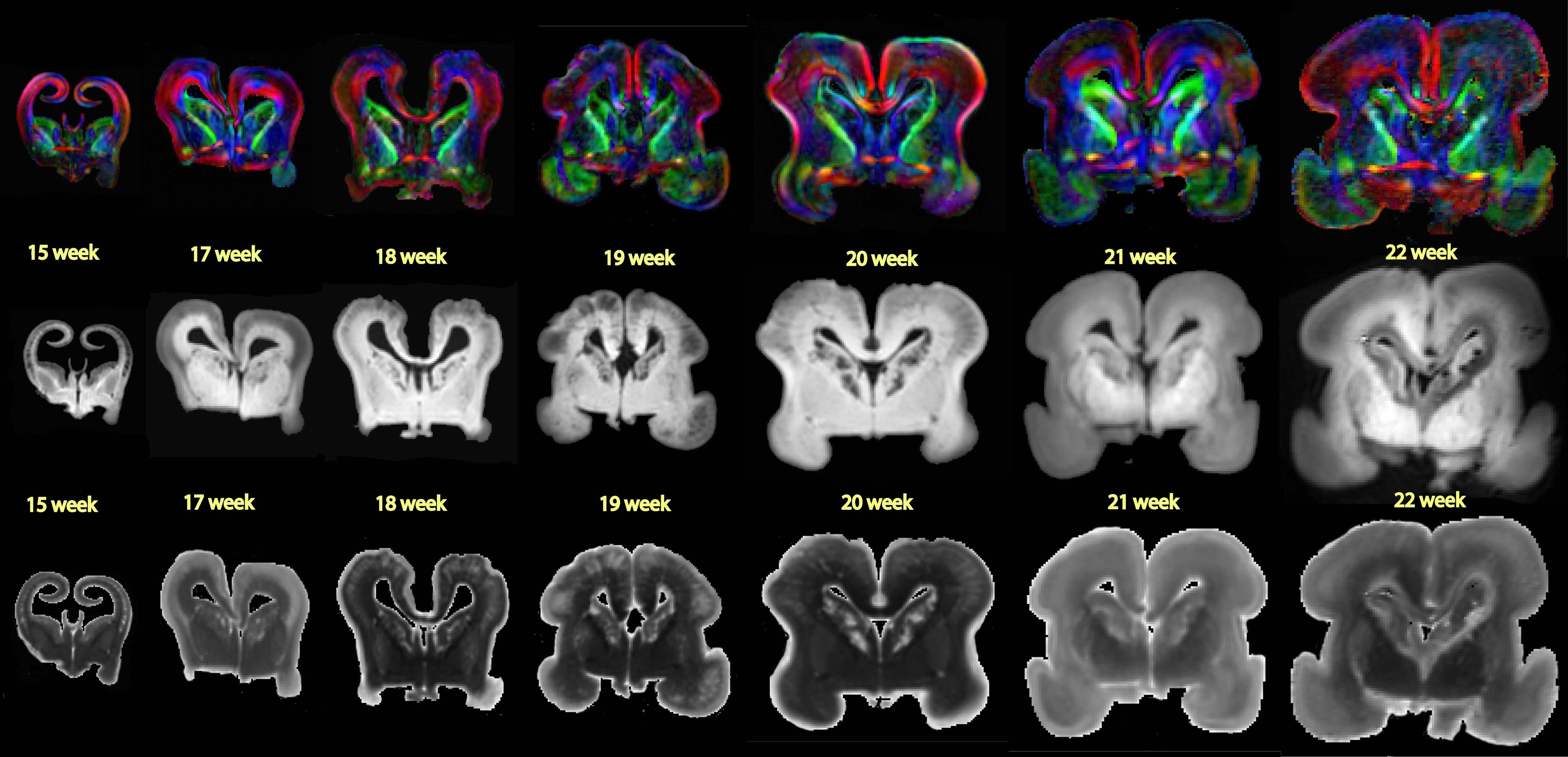
What analytical model is used to estimate critical conditions at the onset of folding in the brain?

The analytical model used to estimate critical conditions at the onset of folding in the brain is based on the Föppl–von Kármán theory. This theory is applied to approximate cortical folding as the instability problem of a confined, layered medium subjected to growth-induced compression. The model focuses on predicting the critical time, pressure, and wavelength at the onset of folding in the brain's surface morphology.


The analytical model adopts the classical fourth-order plate equation to model the cortical deflection. This equation considers parameters such as cortical thickness, stiffness, growth, and external loading to analyze the behavior of the brain tissue during the folding process. By utilizing the Föppl–von Kármán theory and the plate equation, researchers can derive analytical estimates for the critical conditions that lead to the initiation of folding in the brain.


Analytical modeling provides a quick initial insight into the critical conditions at the onset of folding, allowing researchers to understand the fundamental mechanisms driving cortical folding. However, it may not fully capture the evolution of complex instability patterns in the post-critical regime. Therefore, while the analytical model helps in estimating the critical parameters for folding initiation, a computational model based on the continuum theory of finite growth is often employed to predict more realistic surface morphologies and complex folding patterns beyond the onset of folding.


In conclusion, the analytical model based on the Föppl–von Kármán theory provides a foundational framework for understanding the critical conditions that trigger folding in the brain's surface morphology. It serves as a valuable tool for estimating key parameters at the onset of cortical folding and guiding further computational modeling efforts to explore the evolution of brain surface morphologies.


Comments

Popular posts from this blog

Psychoactive Drugs in Brain Development

Psychoactive drugs can have significant effects on brain development, altering neural structure, function, and behavior. Here is an overview of the impact of psychoactive drugs on brain development: 1.      Neuronal Structure : o   Exposure to psychoactive drugs, including alcohol, nicotine, benzodiazepines, and antidepressants, can lead to structural changes in the brain, affecting neuronal morphology, dendritic arborization, and synaptic connectivity. o     Chronic administration of psychoactive drugs during critical periods of brain development can disrupt normal neurodevelopmental processes, leading to aberrations in dendritic spines, synaptic plasticity, and neuronal architecture. 2.      Cognitive and Motor Behaviors : o     Prenatal exposure to psychoactive drugs has been associated with cognitive impairments, motor deficits, and behavioral abnormalities in both animal models and human studies. o  ...

Globus Pallidus Pars Interna (GPi)

The Globus Pallidus Pars Interna (GPi) is a vital component of the basal ganglia, a group of subcortical nuclei involved in motor control, cognition, and emotion regulation. Here is an overview of the GPi and its functions: 1.       Location : o The GPi is one of the two segments of the globus pallidus, with the other segment being the Globus Pallidus Pars Externa (GPe). o It is located adjacent to the GPe and is part of the indirect and direct pathways of the basal ganglia circuitry. 2.      Structure : o The GPi consists of densely packed neurons that are primarily GABAergic, meaning they release the inhibitory neurotransmitter gamma-aminobutyric acid (GABA). o   Neurons in the GPi play a crucial role in regulating motor output and cognitive functions through their inhibitory projections. 3.      Function : o Inhibition of Thalamus : The GPi is a key output nucleus of the basal ganglia that exerts inhibitory control...

Intermittent Theta Burst Stimulation (iTBS)

Intermittent Theta Burst Stimulation (iTBS) is a specific pattern of transcranial magnetic stimulation (TMS) that has gained attention in neuroscience research and clinical applications. Here is an overview of Intermittent Theta Burst Stimulation and its significance: 1.       Definition : o    Intermittent Theta Burst Stimulation (iTBS) is a form of repetitive TMS that delivers bursts of high-frequency magnetic pulses in a specific pattern to modulate cortical excitability. o    iTBS involves short bursts of TMS pulses (burst frequency: 50 Hz) repeated at theta frequency (5 Hz), with intermittent pauses between bursts. 2.      Stimulation Protocol : o    The typical iTBS protocol consists of bursts of three pulses at 50 Hz repeated every 200 milliseconds (5 Hz) for a total of 600 pulses over a session. o    The stimulation pattern is designed to induce long-term potentiation (LTP)-like effects on synap...

How can EEG findings help in diagnosing neurological disorders?

EEG findings play a crucial role in diagnosing various neurological disorders by providing valuable information about the brain's electrical activity. Here are some ways EEG findings can aid in the diagnosis of neurological disorders: 1. Epilepsy Diagnosis : EEG is considered the gold standard for diagnosing epilepsy. It can detect abnormal electrical discharges in the brain that are characteristic of seizures. The presence of interictal epileptiform discharges (IEDs) on EEG can support the diagnosis of epilepsy. Additionally, EEG can help classify seizure types, localize seizure onset zones, guide treatment decisions, and assess response to therapy. 2. Status Epilepticus (SE) Detection : EEG is essential in diagnosing status epilepticus, especially nonconvulsive SE, where clinical signs may be subtle or absent. Continuous EEG monitoring can detect ongoing seizure activity in patients with altered mental status, helping differentiate nonconvulsive SE from other conditions. 3. Encep...

Dorsolateral Prefrontal Cortex (DLPFC)

The Dorsolateral Prefrontal Cortex (DLPFC) is a region of the brain located in the frontal lobe, specifically in the lateral and upper parts of the prefrontal cortex. Here is an overview of the DLPFC and its functions: 1.       Anatomy : o    Location : The DLPFC is situated in the frontal lobes of the brain, bilaterally on the sides of the forehead. It is part of the prefrontal cortex, which plays a crucial role in higher cognitive functions and executive control. o    Connections : The DLPFC is extensively connected to other brain regions, including the parietal cortex, temporal cortex, limbic system, and subcortical structures. These connections enable the DLPFC to integrate information from various brain regions and regulate cognitive processes. 2.      Functions : o    Executive Functions : The DLPFC is involved in executive functions such as working memory, cognitive flexibility, planning, decision-making, ...