
Unveiling Hidden Neural Codes: SIMPL – A Scalable and Fast Approach for Optimizing Latent Variables and Tuning Curves in Neural Population Data

This research paper presents SIMPL (Scalable Iterative Maximization of Population-coded Latents), a novel, computationally efficient algorithm designed to refine the estimation of latent variables and tuning curves from neural population activity. Latent variables in neural data represent essential low-dimensional quantities encoding behavioral or cognitive states, which neuroscientists seek to identify to understand brain computations better. Background and Motivation Traditional approaches commonly assume the observed behavioral variable as the latent neural code. However, this assumption can lead to inaccuracies because neural activity sometimes encodes internal cognitive states differing subtly from observable behavior (e.g., anticipation, mental simulation). Existing latent variable models face challenges such as high computational cost, poor scalability to large datasets, limited expressiveness of tuning models, or difficulties interpreting complex neural network-based functio...

Pivot Joints

Pivot joints are a type of synovial joint that allows rotational movement around a single axis. These joints are crucial for specific movements that involve rotation without significant displacement. Here is an overview of pivot joints:

Pivot Joints:

1.    Structure:

o    Pivot joints consist of a rounded or pointed surface of one bone fitting into a ring or sleeve of another bone or ligament.

o    The structure allows for rotation around a central axis without significant translation.

2.    Function:

o    Pivot joints primarily facilitate rotational movement around a single axis.

o    They provide stability and support for movements that involve twisting or turning.

3.    Examples:

o    Atlantoaxial Joint:

§  The joint between the first (atlas) and second (axis) cervical vertebrae is a classic example of a pivot joint.

§  The dens (odontoid process) of the axis rotates within the ring of the atlas, allowing for rotation of the head.

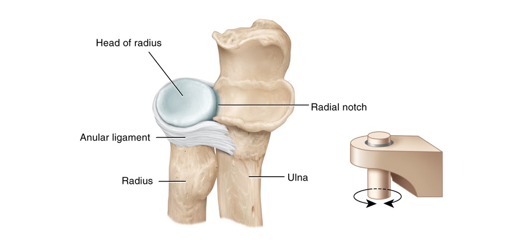
o    Proximal Radioulnar Joint:

§  The joint between the head of the radius and the radial notch of the ulna is another example of a pivot joint.

§  This joint allows for rotation of the radius around the ulna, contributing to movements like pronation and supination of the forearm.

4.    Movements:

o    Rotation: The primary movement at pivot joints is rotation around a central axis.

o    Pronation: Rotational movement that turns the palm downward or backward.

o    Supination: Rotational movement that turns the palm upward or forward.

5.    Stability:

o    Pivot joints provide stability during rotational movements.

o    Ligaments and surrounding structures help maintain the alignment of the bones during rotation.

6.    Clinical Significance:

o    Injuries or conditions affecting pivot joints can impact specific activities that require rotational movements.

o    Rehabilitation programs focus on restoring range of motion, strength, and stability in pivot joints after injuries or surgeries.

Understanding the structure and function of pivot joints is essential for healthcare professionals, athletes, and individuals seeking to maintain joint health and optimize movement capabilities. Proper care, exercise, and biomechanical awareness can help preserve the function and longevity of pivot joints in the body.


Comments

Popular posts from this blog

Sliding Filament Theory

The sliding filament theory is a fundamental concept in muscle physiology that explains how muscles generate force and produce movement at the molecular level. Here are key points regarding the sliding filament theory: 1.     Sarcomere Structure : o     The sarcomere is the basic contractile unit of skeletal muscle, consisting of overlapping actin (thin) and myosin (thick) filaments. o     Actin filaments contain binding sites for myosin heads, while myosin filaments have ATPase activity and cross-bridge binding sites. 2.     Muscle Contraction Process : o     Muscle contraction occurs when myosin heads bind to actin filaments, forming cross-bridges. o     The cross-bridges undergo a series of conformational changes powered by ATP hydrolysis, leading to the sliding of actin filaments past myosin filaments. o     This sliding action shortens the sarcomere, resulting in muscle contract...

What analytical model is used to estimate critical conditions at the onset of folding in the brain?

The analytical model used to estimate critical conditions at the onset of folding in the brain is based on the Föppl–von Kármán theory. This theory is applied to approximate cortical folding as the instability problem of a confined, layered medium subjected to growth-induced compression. The model focuses on predicting the critical time, pressure, and wavelength at the onset of folding in the brain's surface morphology. The analytical model adopts the classical fourth-order plate equation to model the cortical deflection. This equation considers parameters such as cortical thickness, stiffness, growth, and external loading to analyze the behavior of the brain tissue during the folding process. By utilizing the Föppl–von Kármán theory and the plate equation, researchers can derive analytical estimates for the critical conditions that lead to the initiation of folding in the brain. Analytical modeling provides a quick initial insight into the critical conditions at the onset of foldi...

What is Connectome?

  A connectome is a comprehensive map of neural connections in the brain, representing the intricate network of structural and functional pathways that facilitate communication between different brain regions. Here are some key points about the concept of a connectome:   1. Definition:    - A connectome is a detailed representation of the wiring diagram of the brain, illustrating the complex network of axonal projections, synaptic connections, and communication pathways between neurons and brain regions.    - The connectome encompasses both the structural connectivity, which refers to the physical links between neurons and brain areas, and the functional connectivity, which reflects the patterns of neural activity and information flow within the brain.   2. Structural Connectome:    - The structural connectome provides a map of the anatomical connections in the brain, showing how neurons are physically linked through axonal projecti...

How Brain Computer Interface is working in the Cognitive Neuroscience

Brain-Computer Interfaces (BCIs) have emerged as a significant area of study within cognitive neuroscience, bridging the gap between neural activity and human-computer interaction. BCIs enable direct communication pathways between the brain and external devices, facilitating various applications, especially for individuals with severe disabilities. 1. Foundation of Cognitive Neuroscience and BCIs Cognitive neuroscience is the interdisciplinary study of the brain's role in cognitive processes, bridging psychology and neuroscience. It seeks to understand how the brain enables mental functions like perception, memory, and decision-making. BCIs capitalize on this understanding by utilizing brain activity to enable control of external devices in real-time. 2. Mechanisms of Brain-Computer Interfaces 2.1 Neural Signal Acquisition BCIs primarily function by acquiring neural signals, usually via non-invasive methods such as Electroencephalography (EEG). Electroencephalography ...

Pontomedullary Reticular Formation (PmRF)

The Pontomedullary Reticular Formation (PMRF) is a complex network of neurons located in the brainstem, specifically in the pontine and medullary regions. Here is an overview of the PMRF: 1.       Anatomy : o The PMRF is part of the reticular formation, a network of interconnected nuclei and pathways that extends throughout the brainstem. It is situated in the pontine and medullary regions, which are important for regulating various physiological functions. o The PMRF is involved in the modulation of motor functions, sensory processing, cardiovascular control, respiratory rhythm, and the sleep-wake cycle. 2.      Function : o Motor Control: The PMRF plays a crucial role in the coordination of voluntary movements and postural control. It receives inputs from higher brain centers and projects to the spinal cord and cranial nerve nuclei to influence motor output. o   Sensory Processing: The PMRF is involved in sensory integration and modula...